
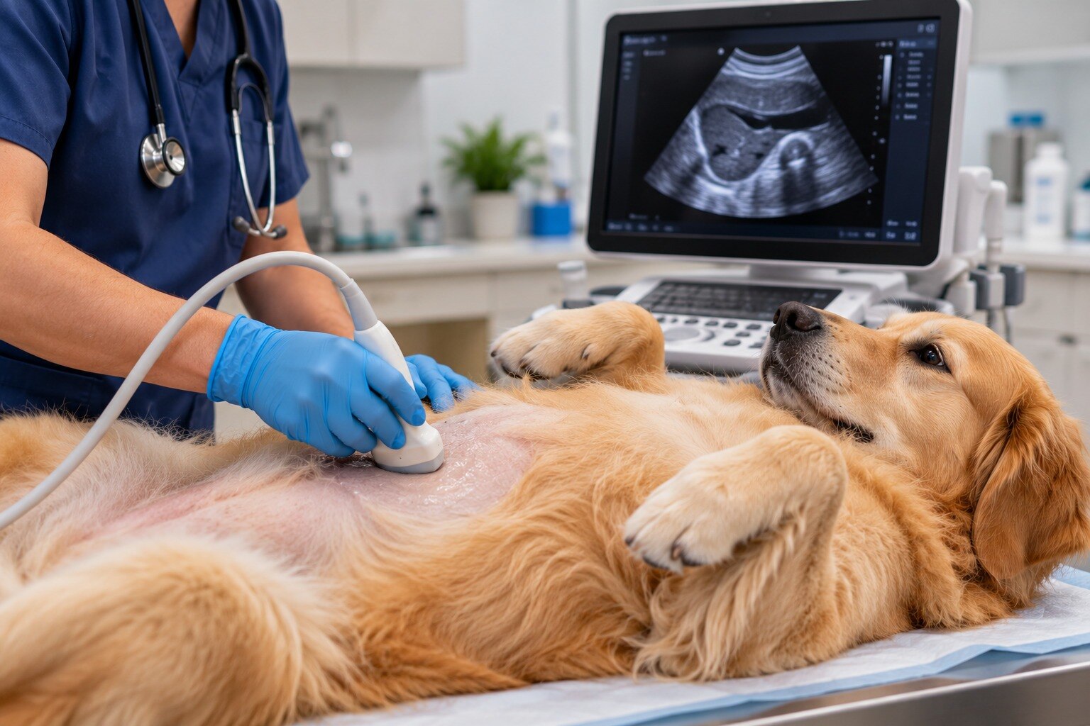
Vomiting, appetite changes, abdominal pain, jaundice, urinary changes, or abnormal liver and kidney results can point to a problem that a physical examination alone cannot locate. By understanding what ultrasound shows, where it falls short, and how the appointment works, you can have a more useful conversation about the next diagnostic step for your pet.
Key takeaways
- Ultrasound shows organ structure, movement, fluid, and Doppler blood flow without radiation.
- Persistent vomiting, abdominal pain, appetite loss, or abnormal blood tests can justify imaging.
- Withhold food only when your veterinarian instructs; fasting improves many abdominal views.
- An abnormal scan usually guides follow-up tests rather than providing a complete diagnosis.
What an abdominal ultrasound shows inside a pet
An abdominal ultrasound for pets is real-time sound-wave imaging of the abdomen. A handheld transducer sends high-frequency sound waves into the body and converts returning echoes into moving images, without ionising radiation. The veterinarian can assess organ size, shape, texture, movement, fluid, and blood flow when Doppler is used.
It can examine:
- Liver, gallbladder, spleen, kidneys, and adrenal glands
- Stomach, intestines, pancreas, and abdominal lymph nodes
- Urinary bladder and the reproductive tract
- Free fluid, cysts, enlarged structures, and many abdominal masses
A liver, gallbladder and spleen ultrasound can reveal changes in organ architecture or fluid around an organ, but an abnormal appearance does not by itself prove cancer, infection, inflammation, or another specific cause. The scan can also guide a needle into selected fluid collections, lymph nodes, or masses for sampling.
Ultrasound and X-rays answer different questions:
| Method | What it shows best | Key limitation |
|---|---|---|
| Ultrasound | Soft-tissue texture, organ layers, fluid, movement, and Doppler blood flow | Intestinal gas, body size, and scanning skill can obscure areas |
| X-ray | Gas patterns, mineralised material, bones, and some foreign objects | Provides less detail about soft-tissue texture and blood flow |
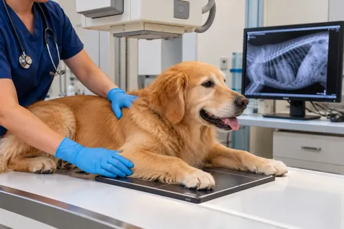
Ultrasound becomes useful when blood-test results or radiographs point to an abdominal problem but do not explain its cause. It does not replace every other scan or test; it adds a detailed, moving view of internal organs.
When abdominal symptoms make ultrasound useful
Persistent or severe symptoms, a painful examination, or abnormal blood or urine results can justify an abdominal ultrasound. It is especially useful when the problem points to soft tissues or an organ but an X-ray cannot explain the cause.
| Sign or test result | Why ultrasound may help | What it may show |
|---|---|---|
| Vomiting and diarrhoea | Looks for intestinal wall changes, obstruction, inflammation, or fluid | Altered intestinal layers, movement, foreign material, or free fluid |
| Abdominal pain and swelling | Examines the abdomen for fluid, enlargement, inflammation, or a mass | Organ changes, enlarged lymph nodes, cysts, or abdominal fluid |
| Unexplained weight loss | Investigates hidden gastrointestinal, pancreatic, liver, or cancer-related disease | Abnormal organ architecture or a mass |
| Appetite changes in pets | Helps assess reduced intake alongside other symptoms or blood-test findings | Stomach, intestines, pancreas, liver, and nearby lymph nodes |
| Jaundice or abnormal liver results | Examines the liver and gallbladder directly | Bile duct blockage, gallbladder changes, or altered liver texture |
| Urinary changes or abnormal kidney results | Evaluates the kidneys and urinary bladder | Stones, obstruction, enlargement, inflammation, or wall changes |
Ultrasound can show that an organ is abnormal without proving whether a mass is cancerous, benign, infectious, or inflammatory. Gas can hide small or early lesions, so the result must be read with the history, physical examination, laboratory tests, and any X-rays or further testing.
How ultrasound compares with X-rays—and where it falls short
Choose ultrasound when you need to inspect soft-tissue structure, fluid, movement, intestinal layers, or Doppler blood flow that a static radiograph cannot show well. It is often the better next test after abnormal blood results or an unclear X-ray, especially when the suspected problem involves an organ rather than bone.
| Test | Best at showing | Choose it when |
|---|---|---|
| Ultrasound | Organ texture, fluid, movement, intestinal layers, and Doppler blood flow | You need soft-tissue detail or guidance for a needle sample |
| X-ray | Gas patterns, mineralised material, bone, and some foreign objects | You suspect an obstruction, swallowed mineral object, fracture, or abnormal gas pattern |
| CT | A broader cross-sectional view of complex masses, gas-obscured areas, and spread to other sites | Ultrasound cannot reach the area or cancer staging requires a more complete survey |
Ultrasound has gas and bone limitations: stomach or intestinal gas can block the sound beam, while ribs and other bones hide structures behind them. Small lesions, early disease, some pancreatic conditions, and some intestinal disorders can also escape detection.
The result depends on positioning, body size, equipment, and examiner training. Ask about referral to a veterinary radiologist or experienced ultrasonographer when findings are subtle, your pet is difficult to scan, or an invasive sample is being considered.
A normal scan does not replace the history, examination, laboratory results, or another scan when those findings remain concerning.
How to prepare your pet for the scan
Ask the hospital for its pet ultrasound preparation instructions before the appointment. A routine scan usually involves placing your pet on a padded table, applying gel to the abdomen, and moving a handheld probe over clipped skin. The images appear immediately on a monitor and often take 20 to 45 minutes.
1. Follow the fasting plan. Fasting before abdominal ultrasound is commonly 8 to 12 hours because food and intestinal gas can hide organs. Confirm whether water and regular medicines are allowed. Do not use the standard fasting period for a diabetic, very young, debilitated, or medically fragile pet without veterinary instructions.
2. Ask about the bladder. A moderately full urinary bladder helps the examiner assess its wall and nearby pelvic tissues, so the hospital may ask you to prevent urination shortly before arrival. Do not force this for a painful, obstructed, incontinent, or unable-to-retain-urine pet.
3. Confirm the restraint plan. Many calm dogs and cats need no anaesthesia. Sedation for ultrasound may be recommended for a fearful, painful, or restless patient, while needle sampling or another invasive procedure can require anaesthesia.
4. Clarify whether this is a complete scan or an emergency check. AFAST can quickly detect free abdominal fluid in a sick or injured pet, but it may not identify the source or replace a full diagnostic examination.
What an abnormal ultrasound means and what may happen next
An abnormal image is a clue, not a diagnosis. Ultrasound findings in pets can reveal an enlarged organ, thickened intestinal wall, fluid, or a suspicious mass, but they rarely prove the underlying cause.
A mass that looks cancerous is not automatically cancer, and a normal scan cannot exclude small lesions, early disease, or problems hidden by intestinal gas.
Definitive diagnosis often requires cells or tissue. Ultrasound-guided fine-needle aspiration can target a lymph node, fluid pocket, organ, or mass while the veterinarian watches the needle in real time. A biopsy after ultrasound provides more tissue, but sampling can miss a lesion or the most important part of a mixed mass.
Bleeding, infection, and injury to nearby structures are weighed against the diagnostic benefit; clotting tests, sedation, or anaesthesia may be needed.
AFAST emergency ultrasound is useful for rapidly checking free abdominal fluid in a seriously ill or injured pet and monitoring changes. It is a screening scan, not a complete abdominal examination, and finding fluid does not identify its source.
Choose the facility by asking whether it offers:
- A complete scan when the problem is not limited to an emergency fluid check
- A veterinarian able to interpret the images alongside examination, blood tests, and X-rays
- Safe sampling, with clotting assessment and appropriate sedation when needed
- A plan for CT, endoscopy, surgery, or oncology referral if ultrasound cannot settle the diagnosis
At Pet Care Hospital- Best Pet Clinic in Dwarka, ask how abnormal findings will be followed through to sampling or referral before booking the scan.
Frequently asked questions
What can an abdominal ultrasound show in a pet?
It can show organ size, shape, texture, movement, abdominal fluid, and blood flow when Doppler is used. It can assess the liver, gallbladder, spleen, kidneys, bladder, stomach, intestines, pancreas, reproductive organs, and nearby lymph nodes.
When might a pet need an abdominal ultrasound?
A veterinarian may recommend it for persistent vomiting or diarrhoea, abdominal pain, appetite or weight loss, jaundice, urinary problems, suspected internal bleeding, abnormal blood results, or a suspected mass.
How is ultrasound different from an X-ray?
Ultrasound creates moving images of soft tissues and blood flow without ionising radiation. X-rays show gas, bone, mineralisation, and some organ size changes more quickly, but they provide less detail for many soft-tissue structures.
How should I prepare my pet for an abdominal ultrasound?
Follow the clinic’s fasting instructions, which improve views by reducing stomach contents and intestinal gas. Ask whether your pet should drink water, take regular medicines, arrive with a full bladder, or have fur clipped from the scan area.
What happens after an abnormal ultrasound?
The veterinarian interprets the finding alongside the examination, history, and blood or urine tests. Follow-up may include needle sampling, biopsy, repeat imaging, endoscopy, surgery, or treatment for the suspected condition.